D is the correct answer
Explanation:
It can be quite stressful working in a single-coverage rural ED with few resources and no in-house consultants, including radiology. Problem-solving and deductive reasoning skills can help decide the best course of action for this patient, given the above chest X-ray.
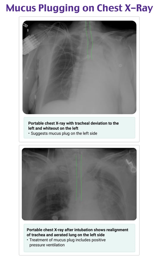
In the image, the trachea clearly deviates to the left. We have all learned that this tracheal deviation means there is a tension pneumothorax pushing the mediastinum to the left. However, no pneumothorax is visible in the image. Furthermore, the left lung whiteout at first glance looks like it could be a large effusion or hemothorax. But it’s not.
When the trachea deviates to the side, it is either due to something pushing it or something pulling it. Since there is no pneumothorax seen on the right, the trachea is not being pushed. Since large effusions generally should not pull the mediastinum toward them, it is unlikely that there is an effusion on the left. Instead, a reasonable conclusion is that there is volume loss in the left lung secondary to airway obstruction that is pulling the trachea toward the left. This is characteristic of a mucus plug that is obstructing inflation of the left lung (similar to all the times your intern right main-stemmed a patient during intubation).
The treatment for this includes positive pressure ventilation noninvasively or through mechanical intubation. Bronchoscopy should be done when available. As you can see below on repeat imaging, this patient was intubated, and positive pressure aerated the left lung, likely breaking up or pushing the mucus plug distally. A chest CT was also ordered on this patient to exclude an empyema or another complication of pneumonia.

References:
- Dixon A. Mucous plug with left lung collapse. Radiopaedia website. Accessed October 19, 2022.
- Friesen B. Left upper lobe collapse due to mucous plugging. Radiopaedia website. Accessed October 19, 2022.
- Panchabhai T, Mukhopadhyay S, Sehgal S, Bandyopadhyay D, Erzurum SC, Mehta AC. Plugs of the air passages: a clinicopathologic review. Chest. 2016;150(5):1141–1157.
P.S. Want to test your knowledge with more questions like this? Take advantage of 15% off the Rosh Review Scholar EM Qbank. You'll get 25 new questions with detailed explanations + images and 50 CME annually!
Use code: RebelEMScholar

