Explanation:
This patient sustained a triquetral fracture. With pain out of proportion after a wrist injury…be aware of the pooping duck sign!
Epidemiology
The triquetrum bone is a pyramid-shaped bone found in the proximal row of carpal bones. Triquetral fractures are the second most common carpal bone fractures, behind the scaphoid, accounting for about 15% of all carpal fractures. These injuries are associated with perilunate dislocations in up to 25% of cases.
The differential includes the following:
- Distal radius and ulna fractures- Extensor carpi ulnaris tendinopathy
- Lunate and perilunate dislocation
- Pisotriquetral arthritis
- Triangular fibrocartilage complex injury
- Wrist sprain
Pathophysiology
The triquetrum bone articulates with three other carpal bones:
- Hamate- Lunate
- Pisiform
These bones protect the triquetrum from direct impact. As a result, triquetral fractures are usually a result of a high-energy impaction by the surrounding bones or ligament avulsion. Over half of these injuries are associated with radiotriquetral ligament tears and > 75% with dorsal scaphotriquetral ligament tears.
The injury mechanism usually involves a fall on an outstretched hand with ulnar deviation and wrist extension.
The pain may seem to be out of proportion. This is due to wrist hyperextension and stretching of the median nerve during a fall.
Remember, axillary nerve injuries occur at the shoulder joint and are seen with humeral head fractures, anterior humeral head dislocations, or improper use of crutches. Radial nerve injuries are seen with midshaft humerus fractures and manifest as weakness with wrist extension, referred to as wrist drop. Ulnar nerve injury would cause fourth (ring) and fifth (pinky) finger paresthesia and weakness of intrinsic hand muscles (weakness of finger spreading and fifth digit flexion and abduction).
Diagnosis
Physical exam can reveal ulnar-sided wrist pain or swelling over the dorsal wrist.
Three-view X-rays are recommended to best visualize a cortical fracture:
- 45° pronated oblique- Lateral
- Posteroanterior
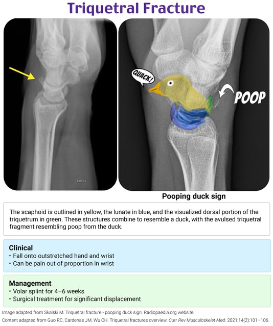
The lateral X-ray is where the pooping duck sign comes in! The term seems to be developed by Radiopaedia. The body of the duck is composed of three bones. The head and neck are formed by the scaphoid bone, the body is composed of the lunate, and the tail is formed by the triquetrum. An avulsed fragment of the dorsal triquetrum appears as if the duck is pooping.
Keep in mind that triquetral fractures can be difficult to visualize on X-rays. X-ray sensitivity is poor, with only 20–30% sensitivity. The pain out of proportion may lead one to reconsider what is being missed with an unremarkable X-ray. CT or MRI would be the next step toward detecting occult fractures and ligamentous injuries.
Treatment
Nonsurgical management with volar splint immobilization for 4–6 weeks is indicated for most triquetral fractures. Thankfully, avascular necrosis is not common with triquetral fractures, unlike scaphoid fractures.
Surgical management with an open reduction and internal fixation is indicated with significant displacement or joint instability.
Complications include the following:
- Nonunion- Pisotriquetral arthritis
- Triangular fibrocartilage complex injury
References:
P.S. Want to test your knowledge with more questions like this? Take advantage of 15% off the Rosh Review Scholar EM Qbank. You'll get 25 new questions with detailed explanations + images and 50 CME annually!

Use code: RebelEMScholar